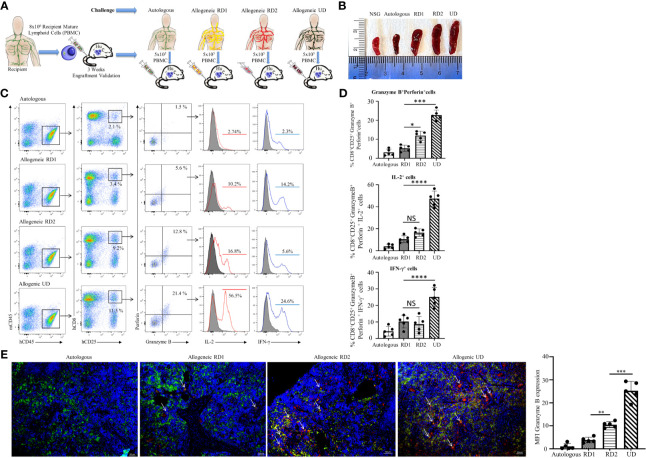
Figure 3.
Hu-NSG-PBMC mouse model exhibits strong CD8+ T cell-mediated allogeneic immune response. (A) Experimental design schematic illustrates humanization of the NSG mouse with PBMCs from the recipient followed by challenge phase with autologous control, allogeneic related donors (RD1, RD2), or allogeneic unrelated donor (UD) PBMCs. (B) Representative spleens isolated from the different challenge groups are shown. (C) Representative flow cytometry color plots depict gating strategy for CD25+ Granzyme B and Perforin expressing activated cytotoxic hCD8+ T cells in spleen of the humanized mouse for the challenge groups. Histogram shows expression of pro-inflammatory cytokines IL-2 (red line) and IFN-γ (blue line) amongst these cytotoxic hCD8+T cells. (D) Graphical summary depicts the frequency (%) of hCD8+CD25+Granzyme B+Perforin+ cells. The IL-2 and IFN-γ profile of CD8+CD25+Granzyme B+ Perforin+ cells in each challenge group is depicted (n=5 mice per group). Data presented as mean ± SD. *p < 0.05, ***p < 0.001, ****p < 0.0001. NS., not significant. (E) Representative immunofluorescent microscopy of spleen sections of the humanized mouse stained with FITC-conjugated hCD8, PE-conjugated hGranzyme B and nuclear staining shown by DAPI (scale bar 5μm). Graphical summary shows mean fluorescence intensity (MFI) for hGranzyme B expression amongst the challenge groups. n=5 mice per group. Data presented as mean ± SD of at least 3 sections from each mouse. **p < 0.01, ***p < 0.001.

