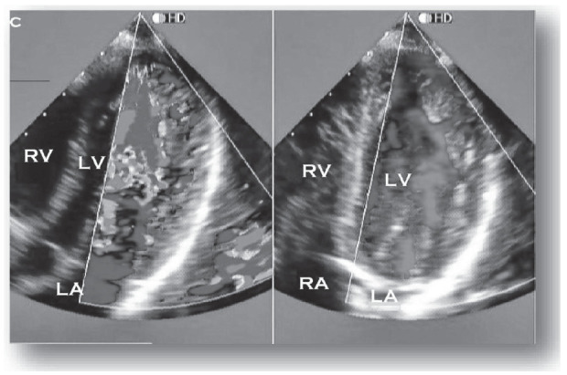
Fig. (1).

Echocardiographic 4-chamber image from a patient with LVNC. The color Doppler highlights perfusion of intertrabecular recesses from the left ventricle. RV: right ventricle; LV: left ventricle; RA: right atrium; LA: left atrium. Courtesy of Paulo G.Menge M.D. (A higher resolution / colour version of this figure is available in the electronic copy of the article).
