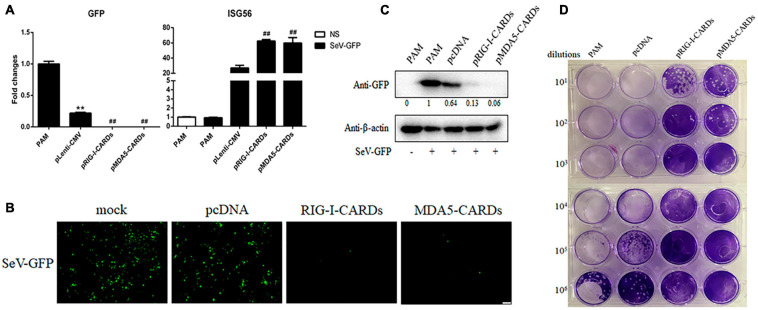
FIGURE 3.
The anti-SeV activity of porcine RIG-I and MDA5 CARDs. (A) PAMs grown on 24-well plates (3 × 10⁵ cells/well) were transfected with pLenti-pRIG-I-CARDs-HA and pLenti-pMDA5-CARDs-HA and pLenti-CMV vector (0.5 μg each) for 24 h. Then the cells were infected with SeV-GFP virus at MOI of 0.01 for 12 h and analyzed by RT-qPCR for virus replication and downstream gene expressions as indicated. (B–D) PAMs grown on 24-well plate were transfected and infected as above. The GFP signals were visualized under microscope (B). The cells samples were detected by Western-blotting with anti-GFP mAb, with the densitometry values after actin normalization shown below the GFP blot. (C). The supernatants from SeV infected PAMs were subjected to infection of Vero cells and the viral plaque were visualized at 24 h post infection (D). **p < 0.01 vs. blank controls; ##p < 0.01 vs. vector controls.

