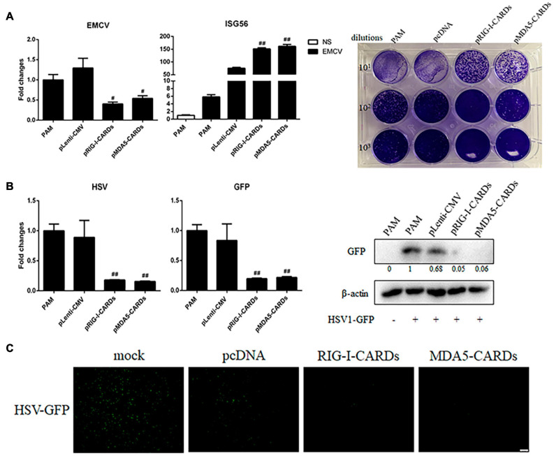
FIGURE 4.
The anti-EMCV and HSV-1 activity of porcine RIG-I and MDA5 CARDs. (A) PAMs grown on 24-well plates (3 × 10⁵ cells/well) were transfected with the indicated CARDs expression plasmids and control vectors (0.5 μg each) using TransIT-LT1 Transfection Reagent for 24 h. Then the cells were infected with EMCV virus at MOI of 0.01 for 12 h and analyzed by RT-qPCR for virus replication, downstream gene expression and by plaque assay for observation of plaque formation in Vero cells at 24 h post infection. (B,C) PAMs grown on 24-well plate (3 × 10⁵ cells/well) were transfected with CARDs expression plasmids and pLenti-CMV vector (0.5 μg each) using TransIT-LT1 Transfection Reagent for 24 h. Then the cells were infected with HSV-1-GFP virus at MOI of 0.01 for 12 h. The cells samples were analyzed by RT-qPCR for virus replication and downstream gene expressions as indicated or detected by Western-blotting with anti-GFP mAb (B). The GFP signals were visualized under microscope (C). #p < 0.05, ##p < 0.01 vs. vector controls.

