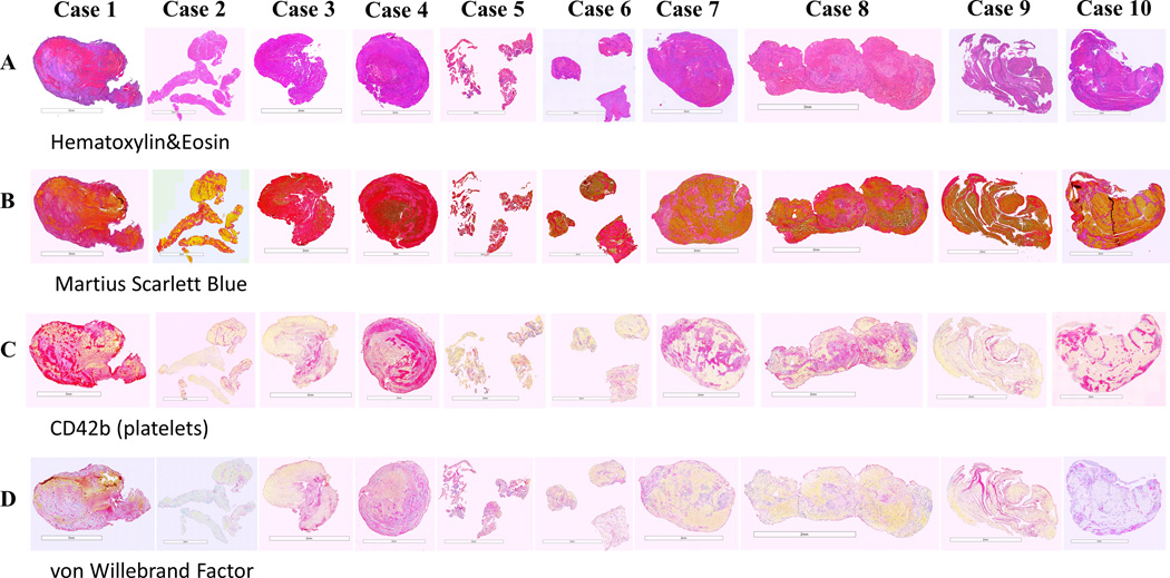
Figure 2. Histological and immunohistochemical analysis of thrombi included in the study.
Consecutive sections were stained with: (A) Hematoxylin and Eosin (H&E), (B) Martius Scarlett Blue (MSB), (C) anti-CD42b (platelets) and (D) anti-von Willebrand factor (vWF) antibodies. H&E shows red blood cells (RBC)-rich areas (red/dark pink) and fibrin/platelets-rich areas (light pink). MSB identifies the main components: RBC (yellow), fibrin (red), platelets/other (light pink) and white blood cells (WBC, blue nuclei). Two distinct morphological patterns were present: 1) an RBC-rich core and fibrin/platelet-rich areas distributed toward the periphery (Case 1 and Case 4); 2) RBC-rich and fibrin/platelet-rich areas interspersed with each other (Case 2, Case 3 and Cases 5–10). WBC were mainly distributed throughout the fibrin-rich and platelet-rich areas. Platelet/other-rich regions identified by MSB stain (B) were also positive (purple) for CD42b (C) and vWF (D). Scale bar = 3 mm (Case 1 and Case 2) and 2 mm (Cases 3–10).

