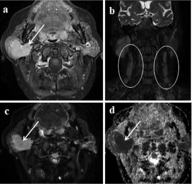
Figure 3.

Lymphoma involvement of the parotid gland 3a: In contrast enhanced axial image; right-sided well-defined enhancing lesion involving almost the entire parotid gland is seen (arrow). 3b: Multiple spheric and oval-shaped lymphadenopathies in the cervical region (circles) 3c, d: Lesion shows marked diffusion restriction (arrows).
