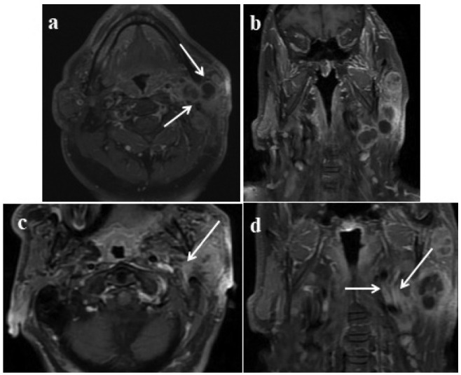
Figure 4.

Multiple lesions in the deep and superficial lobes of the left parotid gland; proven mucoepidermoid carcinoma: Axial 4b: Post-contrast coronal T1W images show left-sided lesions with cystic necrotic areas, heterogenous contrast enhancement in the lesions involving deep and superficial lobes (arrows). 4c: Axial, 4d: T1W post-contrast coronal images; enhancement in the retromolar trigone is adjusted because of perineural spread (arrows).
