Figure 5.
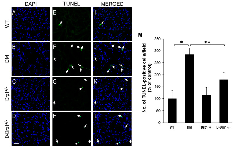
Reduced Drp1 level protects against diabetes-induced apoptosis of vascular cells in retinal capillary networks. Representative images of capillary networks showing DAPI-stained cells in the (A) WT, (B) diabetic (DM), (C) Drp1+/−, and (D) diabetic Drp1+/− (D-Drp1+/−) mice. (E–H) Corresponding images of TUNEL-positive cells (arrows) in the retinal capillary networks, respectively. (I–L) Merged images showing DAPI-stained cells superimposed with TUNEL-positive cells. Scale bar = 100 μm. (M) Graph of cumulative data showing that retinal capillary networks of diabetic mice exhibited an increase in number of TUNEL-positive cells compared to that of WT mice, while retinal capillary networks of D-Drp1+/− mice showed reduced number of TUNEL-positive cells compared to that of diabetic mice. Data are presented as mean ± SD. * p < 0.01, n = 6; ** p < 0.05, n = 6.

