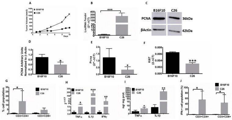
Figure 2.
Reduced tumor growth, immune cell infiltration, and tumor pro-inflammatory cytokine expression correlates with enhanced FENDRR expression. (A) Slower tumor growth rate in C26 compared to B16F10 tumors (n = 5 mice/time point) was noted; (B) A 250-fold enhanced FENDRR expression in C26 versus B16F10 tumors was noted; (C–E) PCNA showed 2–3-fold higher protein and gene expression levels in B16F10 versus C26 tumors. PCNA Western blotting densitometry was normalized to β-actin; (F) KI67 showed 2-fold higher gene expression in B16F10 versus C26 tumors. (G) Enhanced population of CD4+ and CD8+ T cells in C26 compared to B16F10 tumors; (H) Increased expression of TNFα, IL1β, and IFNγ in C26 compared to B16F10 tumors; (I) TNFα and IL1β protein expression levels in C26 tumors were significantly higher than those in B16F10 tumors as determined by ELISA; (J) CD4+ and CD8+ T cells expressed increased IFNγ in C26 compared to B16F10 tumors; Data are shown as mean ± SD. * p < 0.05, ** p < 0.01, *** p < 0.001.

