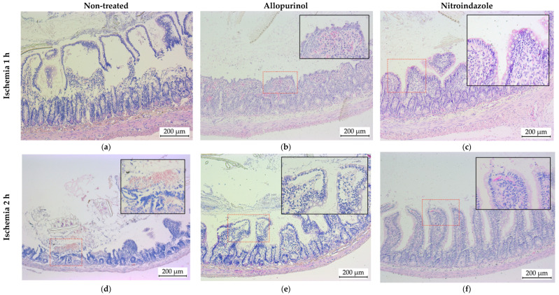
Figure 3.
Representative photomicrographs of small intestine sections stained with hematoxylin/eosin. Animals subjected to a period of 1 (a–c) or 2 h (d–f) of ischemia, followed by 30 min of reperfusion. The photographs on the left (a,c) correspond to histological sections obtained from untreated animals, the photographs in the center (b,d) to animals treated with allopurinol and the photographs on the right (c,f) to animals treated with nitroindazole.

