Figure 2.
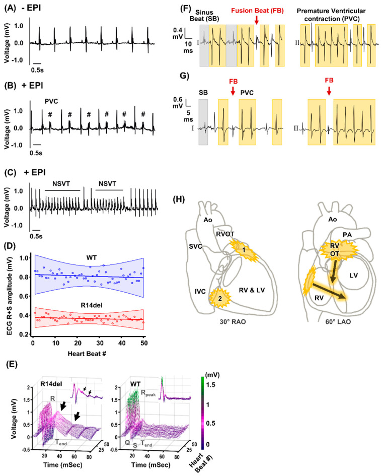
In vivo electrophysiological characteristics and putative source of the ventricular arrhythmias in R14del-PLN mutant mice. (A) ECG tracing under basal condition in R14del-PLN mice. (B,C) ECG tracings showing stress (caffeine and epinephrine) induced arrhythmias in the forms of premature ventricular complexes (PVCs, #), and non-sustained ventricular tachycardia (NSVT) in R14del-PLN mice (N = 5). (D) The mean R+S wave amplitudes with linear regression fit (solid line) and 95% confidence intervals (shaded) are shown for 50 consecutive heartbeats at a steady state for WT and R14del mice. (E) Representative ECG traces are plotted for 50 consecutive heartbeats at a steady state as a function of time and heartbeat number. The R14del mice (left panel) had reduced R wave amplitudes, prolonged QT and increased dispersion of repolarization (arrows) compared to wild type (WT). (F) Representative ECG tracings of PVC and VT beats from R14del-PLN animals are shown from leads I and II. The presence of fusion beats (red arrow) is diagnostic of PVC and non-sustained VT. Compared to the sinus beats (grey shaded areas), the morphology of the PVC and VT beats (yellow) suggests that these beats originated from or near the right ventricular outflow tract (RVOT). (G) Representative example of another common morphology of the PVC and VT beats from leads I and II for another R14del-PLN animal. The net QRS vector suggests these PVC and VT beats originated from the basolateral free wall of RV. (H) The two most common origin sites for PVC and VT in R14del-PLN mice are summarized in the 30° right anterior oblique (RAO) and 60° left anterior oblique (LAO) schematics of the heart (Ao = aorta; SVC = superior vena cava; IVC = inferior vena cava; PA = pulmonary artery; LV = left ventricle).

