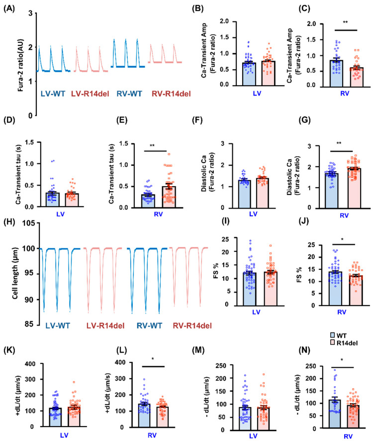
Figure 4.
Ca-kinetic and contractile parameters in isolated cardiomyocytes. (A) Representative traces of Ca-transients; (B,C) Ca transient amplitude, indicated by fura-2 ratio (340:380 nm). (D,E) The relaxation time constant (Tau) of the Ca-transient decay. (F,G) Intracellular diastolic Ca levels in LV and RV myocytes. N = 36 LV and 35 RV cells for WT (N = 4 hearts); n = 34 LV and 41 RV cells for R14del-PLN (N = 4 hearts). (H) Representative cell shortening traces. (I–N) Contractile parameters: Fractional shortening (FS%) and maximum rates of contraction (+dL/dt) and re-lengthening (−dL/dt) in LV and RV myocytes at 0.5 Hz. N = 43 LV and 50 RV cells for WT (N = 4 hearts); n = 40 LV and 43 RV cells for R14del-PLN (N = 4 hearts. Data are expressed as mean ± SEM for the number of cells, and statistical analyses were performed by Student’s unpaired t-test. * p ≤ 0.05 vs. WT-PLN; ** p ≤ 0.001 vs. WT-PLN.

