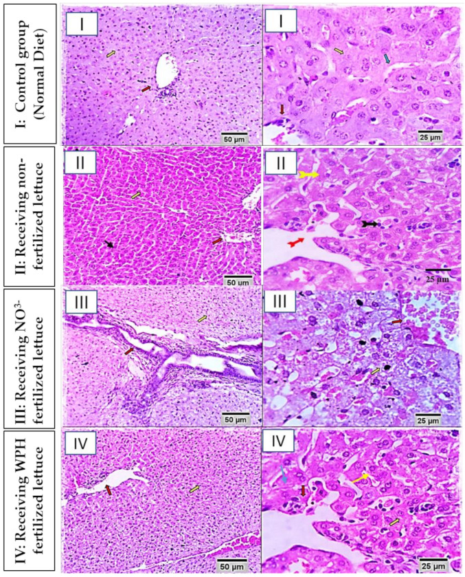
Figure 1.
Photomicrograph from liver of different rabbit groups (I, II, III and IV) at 25 µm and 50 µm scale bars. Group I (healthy control, not receiving any supplement), is showing preserved lobular arrangement, hepatic cords orientations (yellow arrows), portal tirades structural components (red arrows), sinusoids, Von–Kuffer cells (green arrows), and stroma. Group II is showing normal hepatic parenchyma with preserved hepatic cords arrangement (yellow arrows), portal triads structures (red arrows), and sinusoids (black arrow). Group III (rabbits receiving nitrate-fertilized lettuce) is showing moderate portal and interstitial aggregations of round cells, mostly lymphocytes and plasma cells (orange arrow). Bile ducts appear moderately hyperplastic and to be suffering from chronic obstructive cholangitis (red arrow). Moderate portal vascular congestion and sinusoidal dilatation are seen (red and black arrow). Periportal hepatocellular degenerative changes (mostly hydropic degeneration) and early necrotic changes are seen (yellow arrows). Group IV is showing degenerative changes in a few hepatocytes. Most of the degenerative changes were within the cloudy swelling and hydropic types (yellow arrows). Portal tirades showed mild aggregation of lymphocytes and plasma cells (red arrows). Focally dilated hepatic sinusoids are seen (blue arrow).

