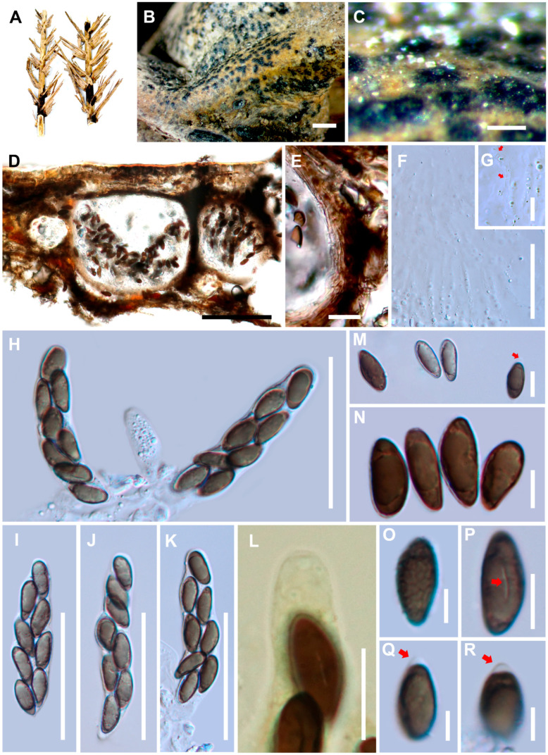
Figure 2.
Haploanthostomella elaeidis (MFLU 20-0522, holotype). (A) Substrate. (B,C) Appearance of ascomata on the host surface. (D) Sections of ascomata. (E) Peridium. (F) Hamathecium. (G) Septa of paraphyses show in red arrows. (H,I–K) Asci. (L) J- apical ring in Melzer’s reagent. (M,N,P–R) Ascospores with mucilaginous cap (red arrows in M, Q, R) and germ slit (red arrows in P). (O) An ascospore with verrucose wall. Scale bars: B = 1000 μm, C = 200 μm, D = 500 μm, E, G, L = 20 μm, F, H–K = 50 μm, M–P = 10 μm, Q–R = 5 μm.

