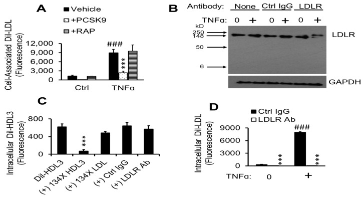
Figure 7.
TNFα-induced Dil-LDL accumulation is blocked by specific LDLR antibody. (A) pHAECs in serum medium were pre-treated with 0 (Ctrl) or 100 ng/mL TNFα for 24 h. Afterwards, the cells were cultured in the presence of vehicle, 12.5 µg/mL human PCSK9 (ApoER, LDLR, LRP, and VLDLR inhibitors), or 21 µg/mL human RAP (ApoER2, LRP, and VLDLR inhibitors) in serum-free medium for 1 h. Lastly, 8 µg/mL Dil-LDL was added, followed by 4 h incubation. n = 3. ###, ***, p < 0.001 vs. Ctrl vehicle and TNFα vehicle, respectively. (B), Cells were cultured in serum medium ± 100 ng/mL TNFα, followed by 3 h incubation in serum-free medium in the continued presence of TNFα (+) without antibody (None), 18 µg/mL control IgG (Ctrl Ig) or LDLR antibody (LDLR), then immunoblotted for LDLR and GAPDH. n = 3. (C), pHAECs were cultured in serum-free medium for 2 h, followed by incubation with 5 µg/mL Dil-HDL3 with (+) 134× unlabeled HDL3, LDL, 20 µg/mL control IgG (Ctrl IgG) or LDLR antibody (LDLR Ab), as indicated. n = 6. ***, p < 0.001 vs. Dil-HDL3. (D) Cells were cultured in serum medium with 0 or 100 ng/mL TNFα (+), followed by 2 h incubation in serum-free medium in the continued presence of TNFα. Subsequently, 5 µg/mL Dil-LDL in the presence of 20 µg/mL control IgG (Ctrl IgG) or LDLR antibody (LDLR Ab) was added. Note that the values for LDLR Ab are very low compared to others. n = 8. ###, ***, p < 0.001 vs. corresponding 0 µg/mL TNFα and Ctrl IgG, respectively.

