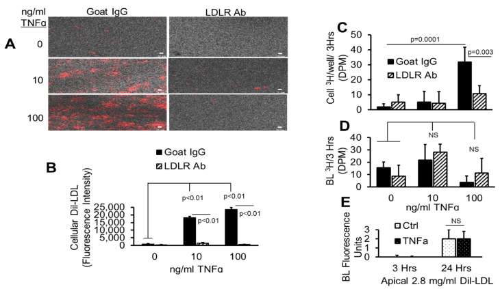
Figure 12.
TNFα does not affect AP to BL LDL lipid release. (A–D) pHAECs were pre-treated as in Figure 11 with 0, 10, or 100 ng/mL TNFα for 24 h in serum. Subsequently, 20 µg/mL control normal goat IgG (Goat IgG) or LDLR antibody (LDLR Ab) was added to the AP medium without serum for 2 h. Finally, 1.6 mg/mL double labeled Dil, [3H]CE-LDL was added to the AP medium for 3 h. After heparin wash, the intracellular fluorescence (A,B), 3H radioactivity (C), or medium BL 3H (D) was determined. (E) The cells were treated for 3 or 24 h with 0 (Ctrl) or 100 ng/mL TNFα without serum in the presence of apical Dil-LDL, and the BL fluorescence was determined. n = 3.

