Figure 3.
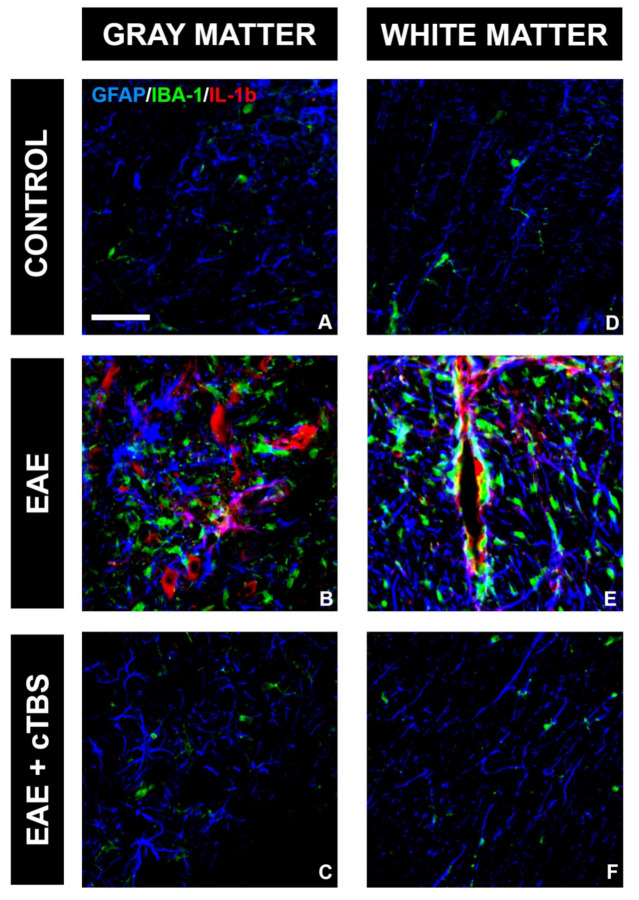
Effect of CTBS treatment on IL-1β expression in gray and white matter of EAE rats. Triple immunofluorescence labeling directed to astrocyte marker GFAP (blue), microglial marker IBA-1 (green), and pro-inflammatory cytokine IL-1β (red). Expression of IL-1β was not detected in control sections (A,D). In EAE sections, increased IL-1β immunostaining in gray (B) and white matter (E), colocalizing with both GFAP and IBA-1 cells. After CTBS treatment, no IL-1β-ir was observed (C,F). Scale bar corresponds to 50 μm.

