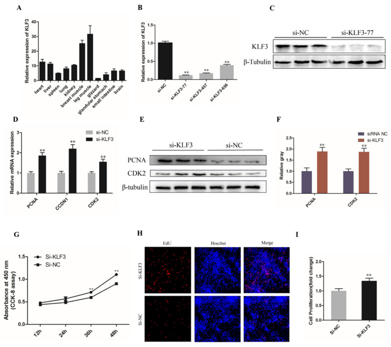
Figure 5.
Knockdown of KLF3 facilitates chicken SMSCs proliferation. (A) Relative expression of KLF3 in different chicken tissues. (B) The knockdown efficiency of KLF3 gene in SMSCs by three small siRNAs were detected by qRT-PCR. (C) The protein expression level of KLF3 after interference by si-KLF3-77 was detected by Western blotting. (D) The mRNA expression of CDK2, PCNA, and CCND1 after 24 h of KLF3 knockdown. (E) CCK-8 assays for SMSCs after KLF3 knockdown. (F,G) The protein expressions of MyHC and MyoG after 48h of inhibition of the KLF3 gene in SMSCs using Western Blot. (H) Results of EdU assay for SMSCs after inhibition of KLF3 for 48 h, where EdU (red) fluorescence is used as an indicator of proliferation and nuclei are indicated by Hochest (blue) fluorescence. (I) The quantitative data of proliferating PSCs number in panel G. The results were expressed as mean ± SEM. (n = 3). ** p < 0.01.

