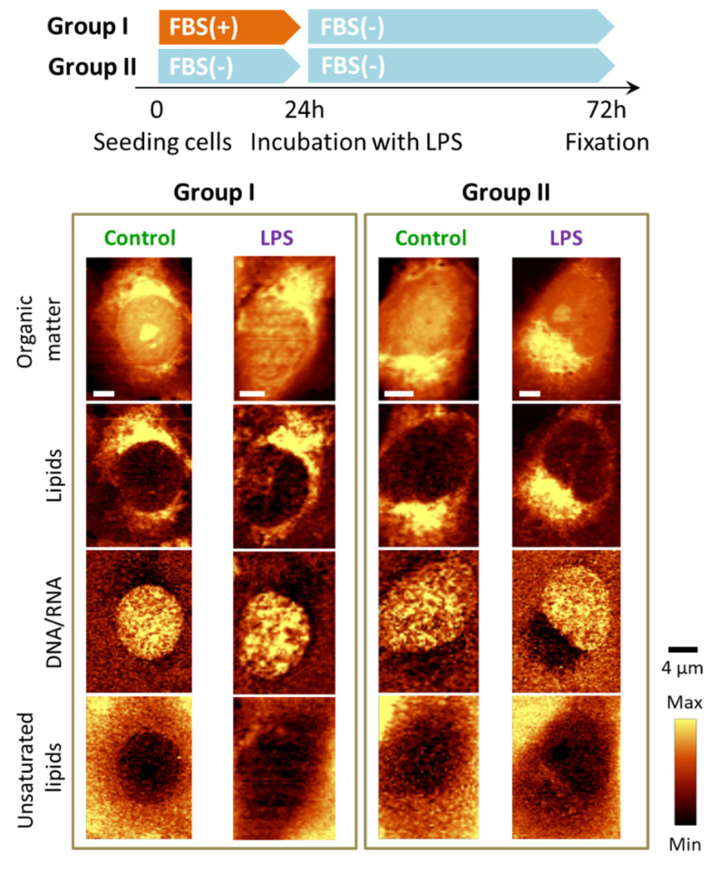
Figure 5.
LPS-induced LDs formation in endothelial cells in the absence of FBS. Representative Raman images of the distribution of HMEC-1 incubated without FBS supplementation in various seeding conditions (absence/presence of LPS) obtained by integration in the following spectral regions: 3030–2830 cm−1 (all organic matter), 2900−2830 cm−1 (lipids), 810−760 cm−1 (DNA & RNA) and 3030–3000 cm−1 (unsaturated lipids).

