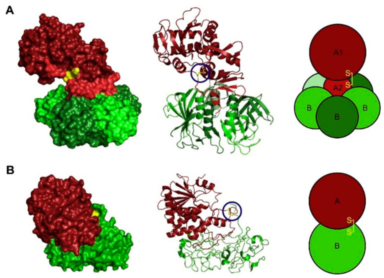
Figure 1.
The structure of (A) Shiga toxin (PDB ID:1DM0 [4] and (B) ricin (PDB ID: 2AAI) [5] determined by X-ray crystallography. The enzymatically active A moieties are colored red, and the B moieties are colored green. The A1 fragment of Shiga toxin is a darker red than the A2 fragment. The disulfide bridge linking the enzymatically active part to the rest of the toxin is indicated in yellow and marked with blue circles in the ribbon structure. Reprinted with permission from ref. [6] Copyright 2014 Elsevier.

