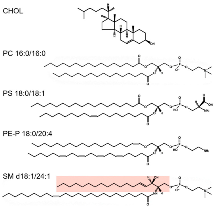
Figure 5.
Illustrations of some lipid structures. On the top, cholesterol (CHOL) is shown, followed by PC 16:0/16:0, which is a lipid species often used in model membranes. However, although PC species are very common in cells, PLs normally contain very little of species with two saturated fatty acyl groups. PS 18:0/18:1 is an example of a PL with 1 saturated and 1 unsaturated fatty acyl group, which is the most common combination of fatty acyl groups in all PL classes. Note that all double bonds in PLs are in a cis-configuration and that the unsaturated fatty acyl group is most often found in the sn-2 position. PE-P 18:0/20:4 is an example of a plasmalogen, i.e., an ether lipid with an alkenyl group, and these lipids often contain polyunsaturated fatty acyl groups in the sn-2 position. The sphingolipid SM d18:1/24:1 is shown with the sphingosine part marked in pink. Note that the N-amidated fatty acyl group is so long that it can theoretically penetrate approximately halfway into the opposite leaflet. These structures were made using the drawing tool available at Lipid Maps (https://www.lipidmaps.org/ (accessed on 24 May 2021)). Reprinted from the open-access review [132].

