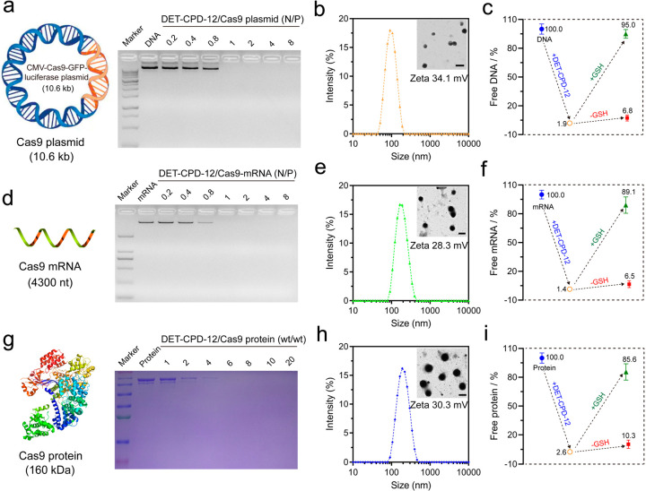
Figure 3.
Preparation and characterization of DET-CPD-12 polyplexes. Agarose gel electrophoresis assay of DET-CPD-12 polyplexes with Cas9 plasmid (a) and Cas9 mRNA (d) at different N/P ratios. Particle size and ζ potential of DET-CPD-12/Cas9 plasmid complexes (b), DET-CPD-12/Cas9 mRNA complexes (e), and DET-CPD-12/Cas9 protein complexes (h). The inset is the TEM image of the nanoparticles, and all of the scale bars represent 200 nm. Percentage of free genome-editing biomacromolecules after the formation of DET-CPD-12/Cas9 DNA complexes (c), DET-CPD-12/Cas9 mRNA complexes (f), and DET-CPD-12/Cas9 protein complexes (i). The release of Cas9 DNA, mRNA, and protein from their complexes was also evaluated in the presence or absence of 10 mM GSH. All data represent mean ± S.D. (n = 3). (g) SDS-page assay of DET-CPD-12/Cas9 protein complexes at different weight ratios.

