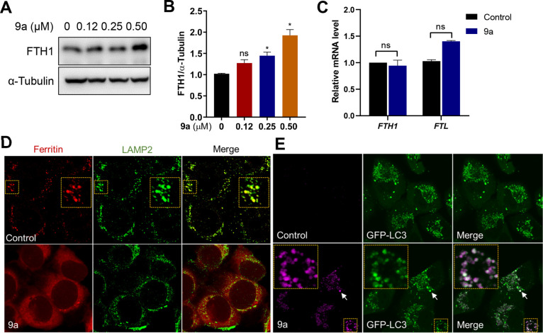
Figure 4.
Compound 9a inhibits lysosomal ferritin degradation and localizes in autophagosomes. (A) Immunoblot analysis of HT22 cells treated with the indicated concentrations of 9a for 6 h. (B) Quantification of the immunoblot analysis in panel A. Data shown represent the mean ± SEM from three independent experiments; *p < 0.05. (C) Effects of 9a on the mRNA levels of FTH1 and FTL. HT22 cells were treated with 0.5 μM 9a for 6 h. Data shown represent the mean ± SEM from three independent experiments. (D) Immunofluorescence imaging of ferritin (red) in HT-1080 cells treated with 9a. LAMP2 (green) stains lysosomes. (E) Confocal imaging of 9a (magenta) in A549 cells that stably expressed GFP-LC3 and were treated for 2 h with the compound at 10 μM. The colocalized foci are indicated by white arrows and shown in an enlarged image of the yellow box.

