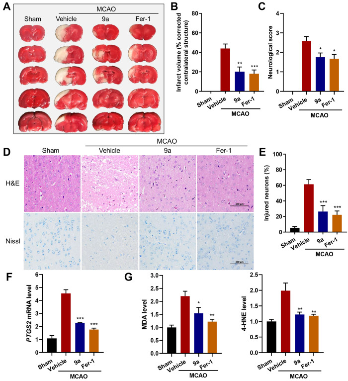
Figure 7.
The administration of 9a ameliorates ischemic reperfusion injury in the MCAO model. (A) Representative images of TTC staining in brain sections at 24 h after MCAO and reperfusion. The infarction area is indicated by the white color with a black outline. Either 9a or Fer-1 was injected into rats 0.5 h before and 2 h after the MCAO occlusion. (B) Quantification of the infarction volume (corrected by the contralateral structure) as indicated by TTC staining using ImageJ (n = 8–9). (C) Statistical analysis of the neurological score (higher numbers indicate more severe impairment) in the Longa test performed at 24 h after MCAO and reperfusion (n = 12). (D) Representative images of H&E (hematoxylin and eosin) and Nissl staining of the cortex of the lesioned hemisphere. The scale bar is 100 μm. (E) Quantification of the percentage of injured neurons in the cortex of the lesioned hemisphere (n = 6). (F) Relative PTGS2 mRNA, (G) MDA, and 4-HNE levels in the lesioned hemisphere (n = 6). Data shown represent the mean ± SEM; *p < 0.05, **p < 0.01, and ***p < 0.001 compared with the group treated with vehicle.

