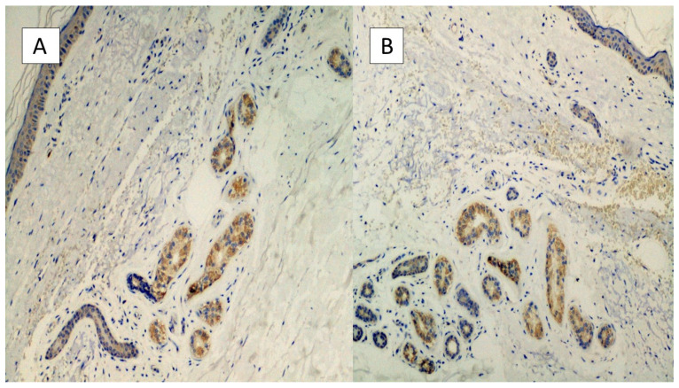
Figure 3.
(A,B) Histological preparation for immunostaining with Anti-SARS-CoV-2 monoclonal antibody. Note the granular, cytoplasmic positivity at the level of the cells constituting the eccrine sweat glands. Involvement of the vascular endothelium is rarely described (IHC, Original Magnification: 10×).

