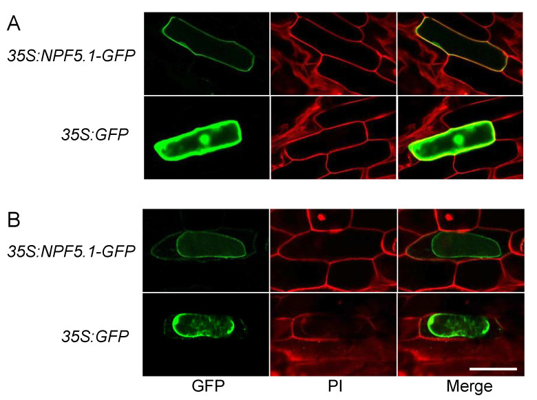
Figure 6.
Plasma membrane localization of NPF5.1. GFP signals in onion epidermal cells that transiently express GFP-fused NPF5.1 or GFP alone under the control of the 35S promoter (35S:NPF5.1-GFP and 35S:GFP, respectively). Photos were taken before (A) and after (B) plasmolysis with 20% mannitol. PI indicates propidium iodide staining of the cell walls. Scale bar = 200 μm.

