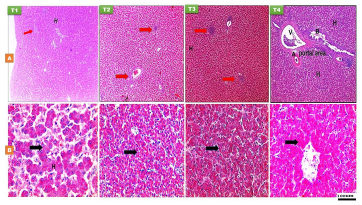
Figure 4.
Histological structure of liver at day 14 (A) and day 28 (B) of age of layer-type chicks exposed to fasting (control, T1) or early feeding on a layer starter diet (T2), a layer starter diet contained 3% molasses (T3), and a broiler starter diet (T4) during the first 72 h post-hatching. Histological examination showed the arrangements of hepatocytes (H) with rounded basophilic nucleus, aggregation of lymph nodules in different area in parenchyma (red arrow) (H and E ×40). Portal area consisted of hepatic portal vein (V), hepatic artery (A), and bile duct (B). The distribution of glycogen granules appeared as reddish dots in the cytoplasm of hepatocytes (black arrow) using Best’s carmine stain (×100 and 200).

