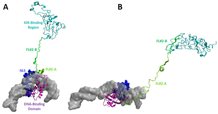
Figure 4.
Stretched linker regions emanating from the DNA-binding and nuclear localization sequence (NLS). The DNA-binding domain is shown in a purple cartoon structure bound to DNA represented as a semitransparent surface. The NLS is shown in dark blue as van der Waals spheres. The first part of Flexible Linker #2 (FOXO3260−321; FL#2-A) is shown as a lime-green cartoon structure, the second part (FOXO3322−344; FL#2-B) in green. The KIX-Binding Region (FOXO3433−508) is represented as a cyan cartoon structure. (A) FL#2-A folds partially in a helical conformation and remains mostly associated with DNA (FL#2-A compact conformation). FL#2-B is highly extended. (B) FL#2-A is present in an extended conformation (FL#2-A extended conformation) and FL#2-B is mostly folded onto the KIX-Binding Region.

