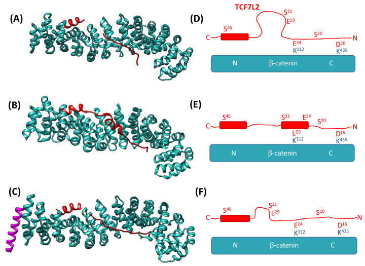
Figure 2.
Schematics showing the crystal structures of the β-catenin–TCF7L2 complex. (A–C) show the structures of TCF7L2 (red) in complex with β-catenin (cyan) obtained by Poy et al. [22], Graham et al. [57], and Sampietro et al. [55], respectively. Structure (C) also has a peptide from the bound protein BCL9 (purple). (D–F) are schematics highlighting the different conformations of TCF7L2 between the three structures and indicating the positioning of the key contact residues within each structure. Images (A–C) were created using UCSF Chimera [24] from PDB 1JPW [22], 1JDH [55], and 2GL7 [25], respectively.

