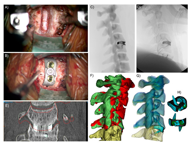
Figure 1.

Stand-alone, integral screw fixation Anterior Cervical Discectomy and Fusion (ACDF) Patient-specific Implant (PSI) C4-5. (A) Surgical discectomy and preparation of the C4-5 interbody space (B) and surgical implantation of the integral screw fixation Titanium alloy PSI. (C) Simulated sagittal plane X-ray used intraoperatively to assess implant positioning (e.g., insertion depth), (D) actual intraoperative sagittal plane X-ray. (E) and three-month postoperative coronal plane CT slice showing fusion bone through the graft window of the Titanium PSI and no discernible subsidence. The red outlines indicate the preoperative position of the C4 vertebra. (F) 3D reconstructions of the cervical levels superior to the operative (C4-5) level; red is the preoperative positioning, and green is the achieved (2.5 month) postoperative positioning. (G) Translucent 3D reconstructions; green is the achieved (2.5 month) postoperative positioning, and blue is the virtual surgery planned (VSP) positioning. Green positioning is close to the matching blue positioning, particularly when compared to red (preoperative) positioning, which shows good surgical realisation of the plan and that anterior interbody devices can control the postoperative segment angle, as well as (height) distraction. (H) Blue (achieved) vs. black (planned) cage positioning within the interbody space; the cage was implanted 0.5–1 mm posterior and to the right of the plan. This was achieved through the use of VSP images (such as C) and as a result of the PSI conforming to the patient’s anatomy, thereby auto-locating in surgery into the planned position.
