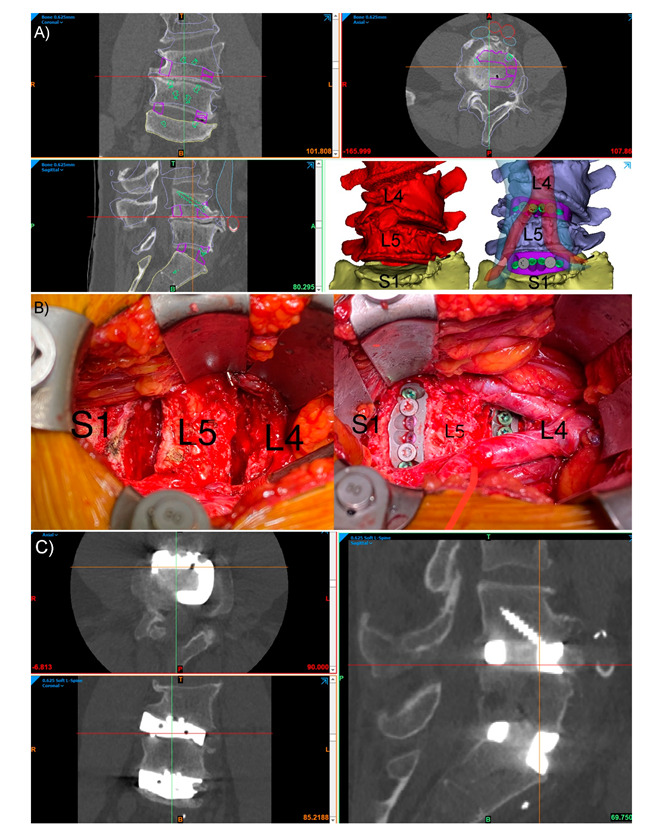
Figure 2.

Integral screw fixation, stand-alone Anterior Lumbar Interbody Fusion (ALIF) patient-specific implants (PSIs) L4-5 and L5-S1, in an L4 congenital hemivertebra patient. (A) Preoperative CT with planned device (purple outlines), screws (green outlines), vertebral positions (blue outlines) and major vessels (inferior vena cava, blue, and aorta, red). The bottom right panel in (A) shows the preoperative pathological anatomy (red) and the planned postoperative state (blue) with translucent aorta (red) and inferior vena cava (blue) shown. (B) The intraoperative L4-L5 and L5-S1 discectomies (left) and final surgical reconstruction (right) with the aortic bifurcation at the L4 level shown. (C) Three-month postoperative CT of the construct showing good positioning of the devices, with no evidence of device migration or micromotion, and interbody fusion bone forming through the graft windows of both PSIs.
