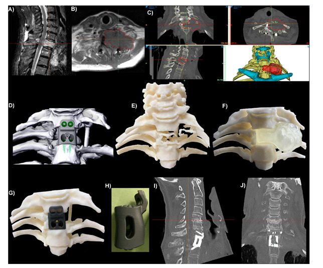
Figure 3.

Integral screw fixation thoracic (T1) corpectomy/vertebrectomy patient-specific implant (PSI). (A) Sagittal plane MRI slice showing a tumour in the T1 vertebral body. (B) Axial plane MRI slice with red outlines showing the tumour. (C) CT slices and 3D reconstruction of the anatomy. The hyoid and sternum are shown in cyan. The position of T1 relative to the sternum meant that access to insert screws up into C7 would be difficult, so a custom anterior plate was integrated into the interbody device with anterior–posterior screw trajectories planned for C7. (D) Virtual Surgical Plan tumour resection and surgical reconstruction using the PSI and integral screws. (E) 3D-Printed ‘biomodel’ of the vertebral and rib bone showing the lytic effects of the tumour on the T1 vertebral and rib bone. (F) 3D-Printed biomodels of the vertebral bone with a removable tumour (opaque, colourless). (G) Same bone biomodel as (F) with the tumour removed and a 3D-Printed resin ‘demo’ PSI in position. (H) Sagittal plane viewpoint of the Titanium alloy (Ti6Al4V) PSI. One-day postoperative sagittal (I) and coronal (J) plane CT slices of the level showing good positioning and contact between the PSI and bone.
