Figure 3.
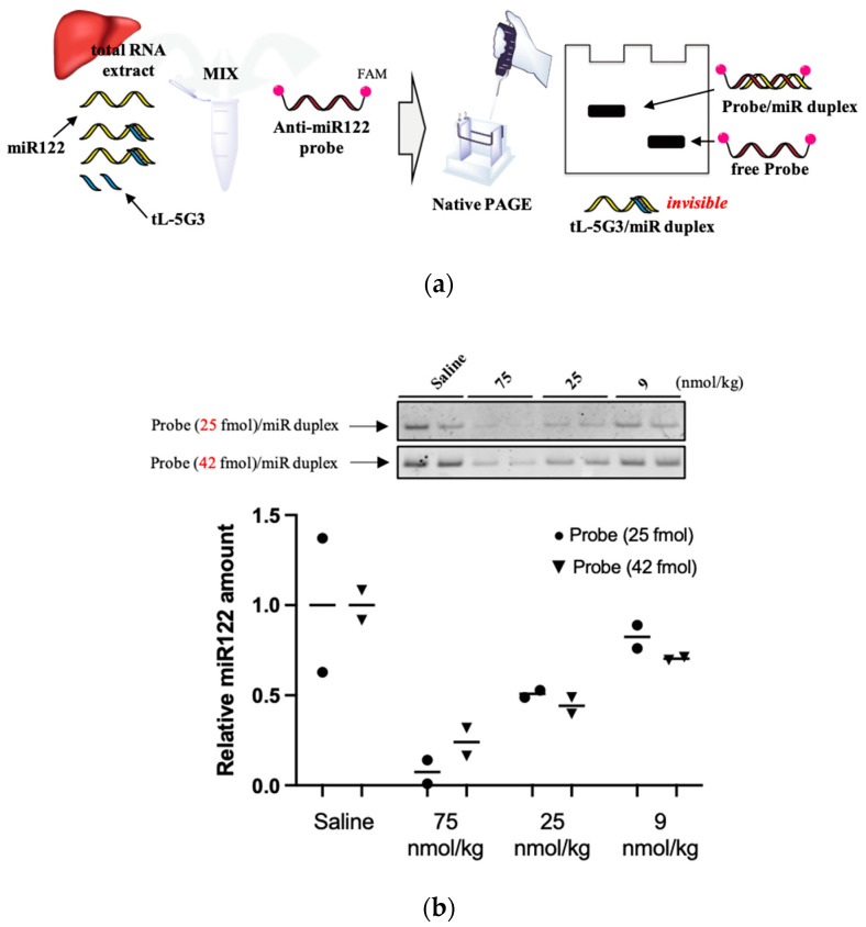
(a) Schematic illustration of the miRNA-122 detection method devised in this study. (b) Representative gel images of the miR122 detection system devised in this study. Total RNA samples obtained from murine liver fragments were analyzed using this detection technique. Total RNA (25 µg) and the FAM-conjugated anti-miR122 probe (25 or 42 fmol) were used in this study. Bands are visualized using Image Quant LAS4000 (GE Healthcare, IL). The fluorescence intensity of the bands was quantified using ImageJ software. The relative amount was estimated from the relative intensity of each duplex band, normalized with the averaged intensity of the duplex bands obtained from each saline-treated arm.

