Figure 2.
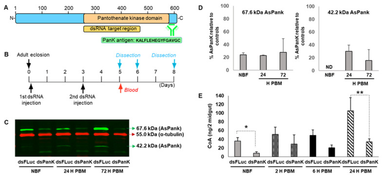
A. stephensi PanK RNAi reduced midgut PanK protein and coenzyme A (CoA) levels. (A). Schematic of A. stephensi PanK indicating the target location of the dsRNA construct and the peptide sequence used to generate the custom polyclonal antibody. (B). Experimental design for RNAi assays. A. stephensi females were injected with dsRNA targeting A. stephensi (dsPanK) or firefly luciferase (dsFLuc) within 4 h after adult eclosion and again at 3 d post-emergence. Mosquitoes were provided human blood on day 5 and midguts were dissected prior to blood feeding (non-blood-fed, NBF) and at 24 h and 72 h post-blood meal (PBM). (C). Representative immunoblot showing putative A. stephensi PanK isoforms (67.6 kDa and 42.2 kDa; green bands) following PanK RNAi relative to FLuc RNAi control (red band). Each lane represents one midgut equivalent from pools of ten mosquito midguts. RNAi and immunoblots were replicated twice with distinct cohorts of mosquitoes. (D). Densitometry analysis of the 67.6 kDa and 42.2 kDa A. stephensi PanK isoforms. Bars represent mean and standard deviation of percent PanK protein expression in mosquitoes inoculated with dsPanK relative to mosquitoes injected with dsFLuc. (E). Midgut CoA levels in dsPanK- and dsFLuc-treated A. stephensi. Differences between treatment groups for respective timepoints were evaluated using Student’s t-test. ** p < 0.01 and * p < 0.05. Experiments were replicated three times with independent cohorts of mosquitoes.

