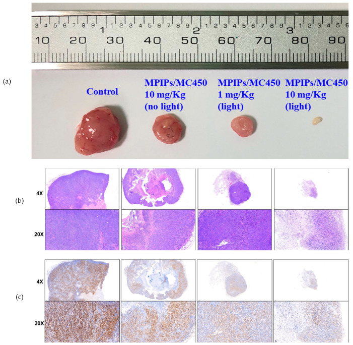
Figure 6.
(a) Pictures of tumor specimens from the xenograft models with treatment of PBS (control), 10.0, 1.0 and 10.0 mg/kg of MPIPs/MC540; the latter two groups were irradiated with 15 min of an LED mini dot light 1 h after injection. (b) Hematoxylin & eosin (HE) staining of above tumor specimens. (c) Immunohistochemical (IHC) staining with anti-proliferating cell nuclear antigen (PCNA) primary antibodies.

