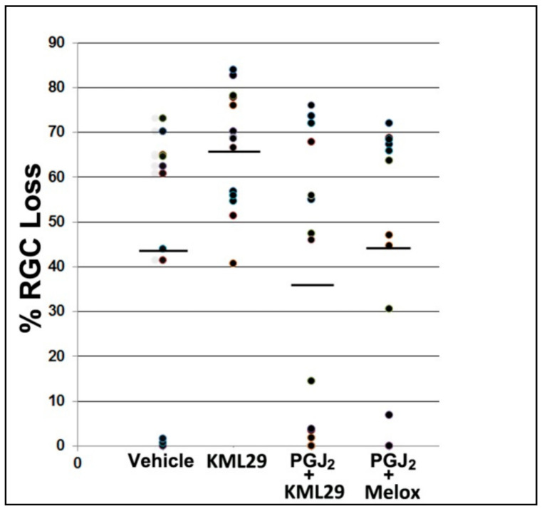
Figure 5.
Stereological analysis of RGC loss following rAION induction and treatment. Scatter plot results of Brn3a(+) flat-mounted RGCs 30 d post-rAION induction. Each point represents the RGC loss from one animal compared with the contralateral (uninduced) eye (=100%). Mean RGC loss in the vehicle group was 44.0 ± 9.8% sem. KML29 treatment alone (no PGJ2 coadministration) resulted in a statistically significant decrease in RGC survival (66.6 ± 3.7% sem; two-tailed t-test; p = 0.0275). There was no statistically significant improvement in RGC survival with PGJ2 + KML29 (mean 36.6 ± 7.9 sem (n = 16) vs. vehicle; 44.0 ± 9.8 sem (n = 11); two-tailed t-test; p = 0.5651), or with PGJ2 + the COX inhibitor meloxicam (Melox) (44.6 ± 9.0 sem (n = 11) vs. vehicle, 44.0 ± 9.8 sem; Mann–Whitney one-tailed test; p = 0.4286), when compared with vehicle alone.

