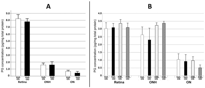
Figure 6.
PGJ2 and PGE2 levels in the retina, optic nerve head (ONH) and optic nerve, and the effects of KML29 treatment (n = 3) on PGE2. White and black bars represent results from naïve (n = 3) and vehicle-injected, 1d post-rAION (n = 3) induced eyes, respectively. (A) PGJ2 levels in retina, ONH and ON. Naïve (Veh OS) retinal PGJ2 levels are considerably higher (8.22 ± 0.51 (sem) than those of ONH (1.58 ± 0.24 (sem) or ON (0.66 ± 0.06 (sem), respectively, and there is little change 1d after rAION induction (Veh OD). (B) PGE2 levels in retina, ONH and ON and the effects of KML29. PGE2 levels in the retina are similar in both naïve (Veh OS) and induced (Veh OD) conditions and KML29 does not significantly alter intraretinal expression at one day post-induction (KML OD). KML OS represents the contralateral (uninduced) ocular tissues. PGE2- expression is slightly less in ONH, and KML29 slightly upregulates PGE2 expression. PGE2 expression is ~2-fold less in the distal ON than in the ONH, and neither rAION nor KML29 significantly affect PGE2 expression in any tissue. Units are in pg/mg protein.

