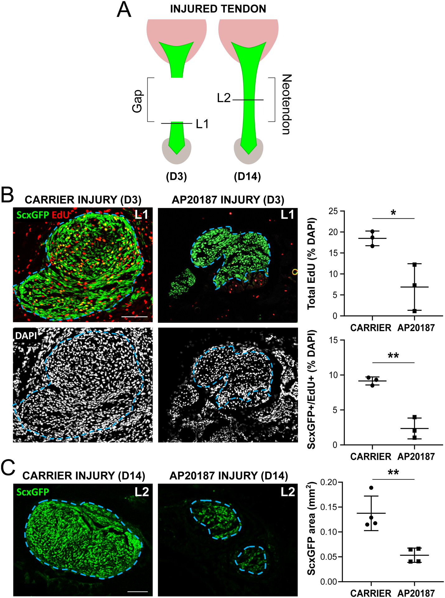
Figure 7: Reduced proliferation and ScxGFP neo-tendon formation after tendon injury with macrophage depletion.

(A) Transverse cryosections were collected from tendon cut site at D3 (L1) and neo-tendon region at D14 (L2) post-injury. (B) EdU and ScxGFP imaging and cell quantification at D3 post-injury (n=3). (C) ScxGFP area quantification at D14 post-injury (n=4). Blue dashed outlines show tendon region of interest based on ScxGFP expression. Data reported as mean±stdev and analyzed by Student’s t-tests. * p<0.05 ** p<0.01. Scalebars: 100 μm.
