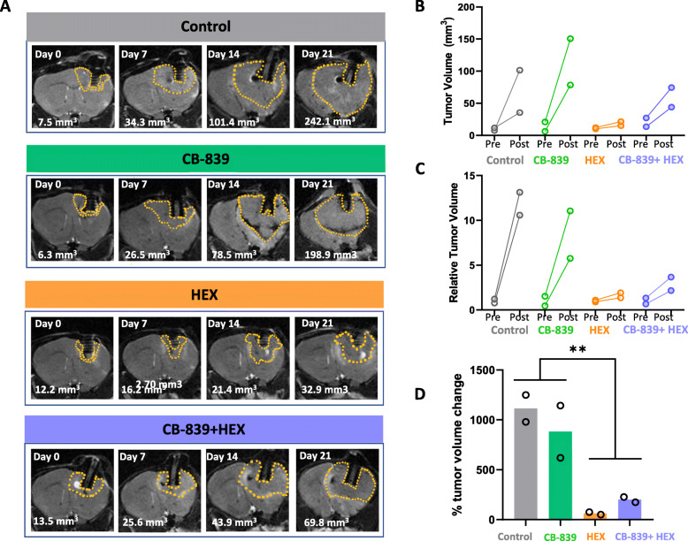
Fig. 5.
CB-839 and HEX combination attenuates intracranial tumor growth but does not cause a frank tumor regression. ENO1-deleted glioma cells (D423) were implanted intracranially in immunocompromised nude mice and tumor growth was monitored weekly by T2 MRI. Tumors are MRI detectable (indicated by dashed yellow outlines) 20-30 days after tumor implantation. 3D slicer was used to view the DICOM files and measure tumor volumes. a Representative MRI images to indicate weekly changes in tumor volume across different treatment groups, control (N=2), CB-839 treated (200 mpk BID orally; N=2), HEX treated (300 mpk SC; N=2), CB-839+HEX (200 mpk CB-839 BID orally, and 300 mpk HEX SC; N=2). b Pre- and post-treatment comparison of absolute and relative tumor volumes across different treatment groups. c Percent change in tumor volume after 2 weeks of drug treatment. Asterisks indicate statistical significance p<0.05 achieved by two-way ANOVA and Tukey’s post hoc analysis. Note that only the effect of HEX is significant

