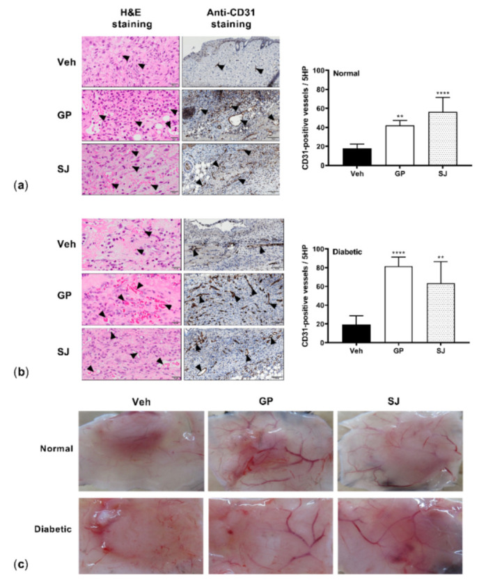
Figure 5.

G. procumbens promotes blood vessel formation: Representative images of wounds from (a) normal and (b) diabetic mice treated with Vaseline (Veh: vehicle), 0.5% G. procumbens (GP) or 10% solcoseryl jelly (SJ). On day 8 post-treatment, sections were histologically stained with H&E (left panels) or immunohistochemically stained with anti-CD31 (middle panels). Scale bar = 20 μm for H&E and 100 μm for anti-CD31 staining. Right panels: the number of CD31-positive vessels. ** p < 0.01 and **** p < 0.0001 for the comparisons between the vehicle and G. procumbens (GP) or solcoseryl jelly (SJ). (c) Representative pictures of the macroscopic appearance of new blood vessels at the wound sites 8 days postinjury on the normal and diabetic mice treated with Vaseline (Veh: vehicle), 0.5% G. procumbens (GP) or 10% solcoseryl jelly (SJ).
