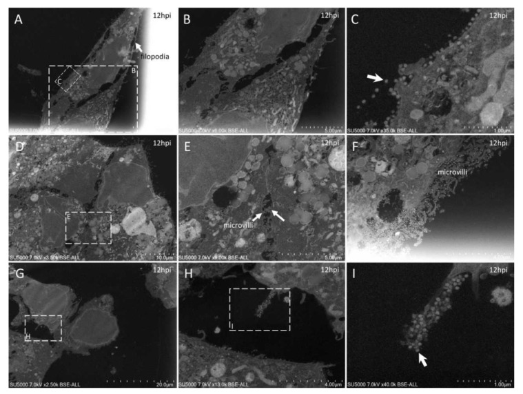
Figure 4.
Ultra-thin sections of SARS-CoV-2-infected Vero E6 cells: 12 hpi. (A–C): Two neighboring cells, one with a long filopodia (A,B; arrow A) and one presenting extracellular SARS-CoV-2 particles at the plasma membrane (C; arrow). (D–F): SARS-CoV-2-enriched microvilli located between neighboring cells (E; arrows) or at the cell-free periphery (F). (G–I): One microvillus extremely enriched in SARS-CoV-2 virions (I; arrow).

