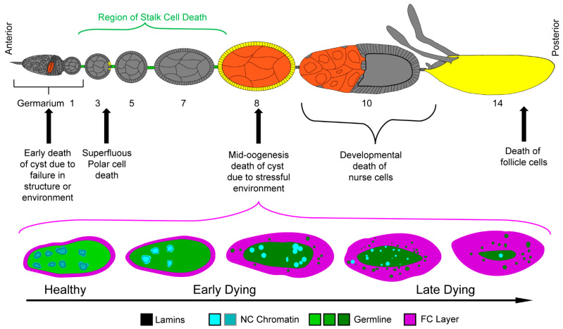
Figure 3.
Cell death in oogenesis. Top—Schematic of ovariole with germline cell death events highlighted in orange and somatic cell death events highlighted in yellow and green. Bottom—Phases of cell death in egg chambers during mid-oogenesis are illustrated. Healthy egg chambers contain NCs with dispersed chromatin and intact nuclear lamina. The FC layer (magenta) surrounding the syncytium is thin, just 1 cell thick. As the germline dies, the chromatin condenses (as shown by the increasing brightness of the cyan) and the lamins are cleaved and become cytoplasmic (as shown by the germline green becoming darker). The chromatin condenses and fragments as germline cell death progresses. The follicle cells then efferocytose the germline material (green vesicles) to clear it away.

