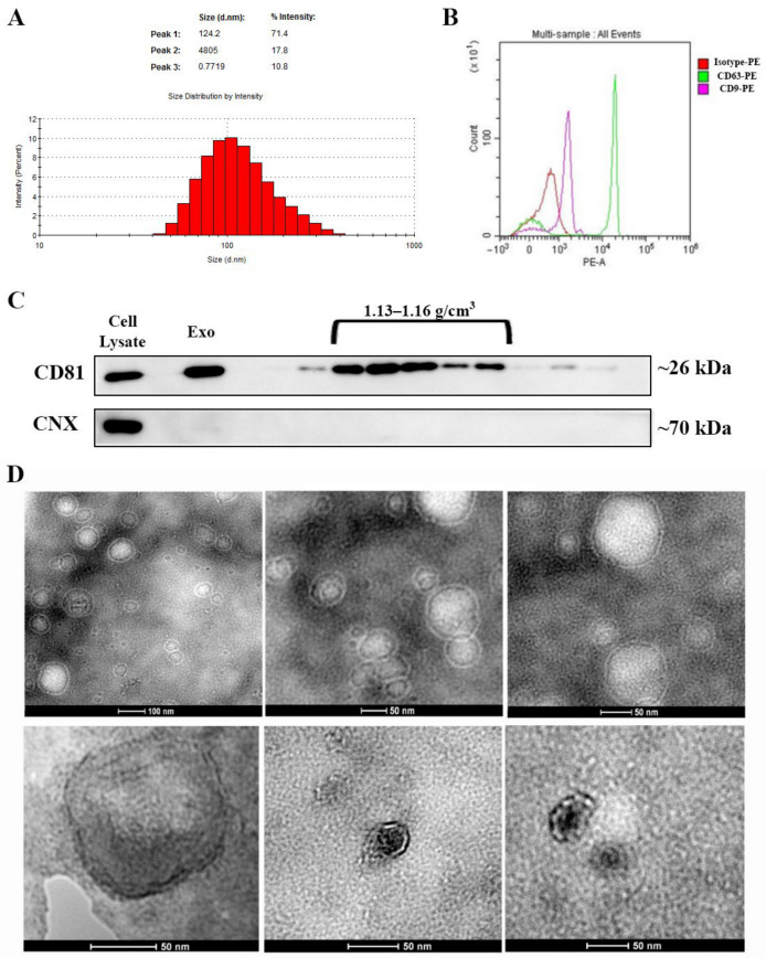
Figure 1.
Characterization of exosomes isolated from bone marrow-derived MSC conditioned medium. (A) Histogram showing the distribution of the hydrodynamic diameter of the isolated exosomes by dynamic light scattering analysis. (B) Expression of exosomal markers CD63 and CD9 as revealed by flow cytometry and (C) CD81 as identified via Western blot for the whole pellet (Exo) as well as for the sucrose fractions derived from it. Note the absence of contaminants from the endoplasmic reticulum as indicated by the lack of calnexin (CNX) both in the exosomes (Exo) pellet and the sucrose fractions. (D) Transmission electron microscopy images depicting donut-shaped structures up to 100 nm (negative staining with phosphotungstic acid—upper panel and uranyl acetate—lower panel).

