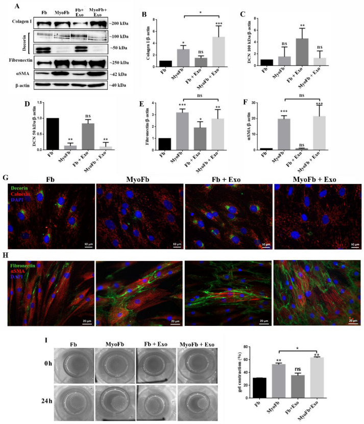
Figure 7.
Exosomes stimulate the expression of proteins involved in matrix remodeling and contraction in fibroblasts differentiated towards myofibroblasts. (A) Representative Western blots images and quantification relative to β-actin for type I collagen (B), decorin (C,D), fibronectin (E), and αSMA (F). (G) Immunocytochemistry image indicating the changes of decorin in fibroblasts treated with TGFβ1, exosomes, TGFβ1 and exosomes, and its distribution in relation to the endoplasmic reticulum evidenced by calnexin. (H) Fluorescence microscopy image showing the organization of αSMA in stress fibers in TGFβ1-treated samples and the extracellular organization of fibronectin. (I) Gel contraction assay demonstrating the contractile activity of fibroblasts as such (control), differentiated (TGFβ1), incubated with exosomes (Exo), and simultaneously stimulated with TGFβ1 and exosomes (TGFβ1 + Exo). The results are given as the percentage of the initial area of the gel. Data are means ± SD (n = 3), * p < 0.05; ** p < 0.01, *** p < 0.001, ns—not significant.

