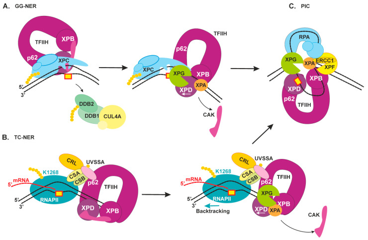
Figure 3.
Schematic view of the damage verification step of NER and pre-incision complex formation. (A) GG-NER. TFIIH initially interacts with XPC’s N terminus by means of p62 subunits, then tumbles to XPC’s C terminus, where the interaction with XPB promotes its binding to a duplex part. XPA releases an inhibitory CAK module and together with XPG stimulates XPD activity [45]. The XPD helicase binds to the damaged strand and starts to a repair bubble formation [7]. When XPD gets to the lesion and stalls on it, XPC is displaced, and XPG binds to the 3′ edge of the repair bubble. (B) TC-NER. XPC and UVSSA share an interaction surface on the p62 subunit of TFIIH [41]. RNAPII moves in the 3′→5′ direction on the damaged strand, then, after its lesion stalling and assembly of factors CSB, CSA, and UVSSA, the latter promotes TFIIH binding downstream of RNAPII [37]. Thereafter, XPA and XPG stimulate XPD activity, and TFIIH starts to move in the 5′→3′ direction and may “push” RNAPII for a backtracking movement. (C) The NER pre-incision complex (PIC): TFIIH stalls on the lesion-bearing strand, RPA covers the undamaged strand, XPA marks the 5′ edge of the repair bubble, XPG marks the 3′ edge of the repair bubble, and XPF–ERCC1 binds behind XPA.

