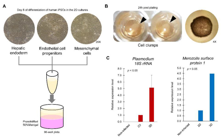
Figure 3.
Potential of liver bud-like cell clumps to model liver-stage malaria. (A) Protocol to generate hepatic endodermal, endothelial and mesenchymal cells from the human iPSC MUi019 line. Cells were harvested from a monolayer of 2D culture and mixed in culture medium. The cell mixture was then added to a presolidified 50% Matrigel (pink) and allowed to settle on the surface. (B) At 24 h of cell plating, the cells had shrunk and became clumped (arrowheads). Microscopic observation revealed the round shape of a cell clump (4× objective lens). (C) At day 6 post inoculation of P. vivax sporozoites, the mRNA expression of Plasmodium 18S rRNA and merozoite surface protein 1 in the 3D culture of liver bud-like cell clumps was higher than that in the 2D culture of human hepatocytes derived from the human iPSC MUi019 line.

