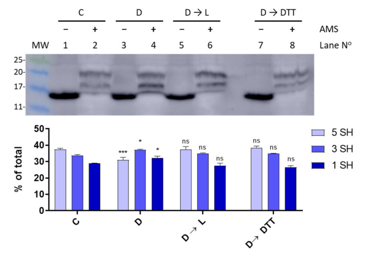
Figure 5.
Light-dark modulation of FurA thiol oxidation. The in vivo redox status of FurA was assessed by Western blot under standard culture conditions of 30 μmol photons m−2 s−1 of white light (C), after 15 min of darkness (D), after 15 min of darkness followed by 3 h of exposure to 30 μmol photons m−2 s−1 of white light (D→L), and after 15 min of darkness followed by 3 h of exposure to 30 μmol photons m−2 s−1 of white light and 2 mM DTT (D→DTT). In all cases, Anabaena sp. PCC 7120 cultures were treated with 10% (w/v) trichloroacetic acid before being alkylated with 20 mM AMS. The relative abundance of each band was quantified by scanning densitometry and represented data are mean ± standard deviation of two biological replicates and two technical replicates. Significance was measured using one-way analysis of variance (ANOVA) comparing with the control (C): ns non significant, * p < 0.05, *** p < 0.00.

