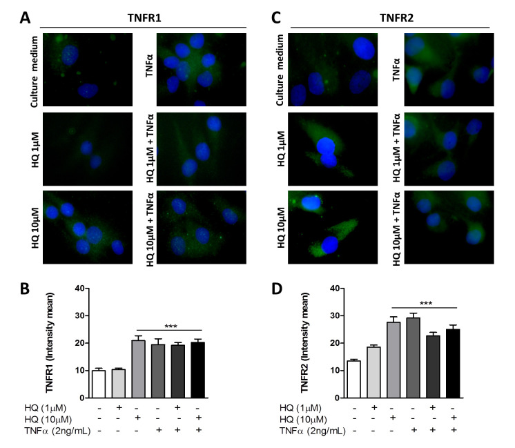
Figure 3.
In vitro HQ treatment induces TNFR1 and TNFR2 expression in RAHFLS. RAHFLS (1 × 10⁴ cells) were incubated with culture medium, or TNF-α (2 ng/mL) or HQ (1 or 10 µM) in presence or absence of TNF-α (2 ng/mL) for 24 h. Then, the expression of TNFR1 (A) and TNFR2 (C) were quantified through an indirect immunofluorescence assay and the mean intensity of immunoreactive areas were quantified (B,D). DAPI—positive staining for nuclei. Original magnification—100×. Data represent mean ± SEM from three independent experiments and were analyzed by one-way ANOVA. *** p < 0.001 vs. culture medium or HQ 1 µM.

