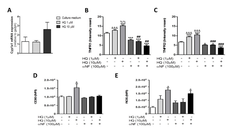
Figure 5.
In vitro AhR antagonist treatment prevents cell proliferation and TNFRs expression evoked by HQ exposure. RAHFLS (1 × 10⁴ cells) were treated with HQ (1 or 10 µM) with or without the AhR antagonist α-naphthoflavone (αNF, 100 µM). After 30 min of treatments, the Cyp1a1 mRNA expression was quantified by RT-PCR (A). After 24 h of treatments, the TNRF1 and TNFR2 expression were quantified by indirect immunofluorescence assay in the synovial cells (B,C) and the synovial proliferation was quantified through flow cytometry (D). The ROS generation was quantified using DCFH-DA assay (E). Data represent mean ± SEM of three independent experiments in RAHFLS. Data were analyzed by one-way ANOVA. * p < 0.05 and *** p < 0.001 vs. culture medium; & p < 0.05 and &&& p < 0.001 vs. αNF; %% p < 0.01 vs. HQ 1 µM; @@ p < 0.01 vs. HQ 10 µM + αNF; ## p < 0.01 and ### p < 0.001 vs. respective groups without αNF.

