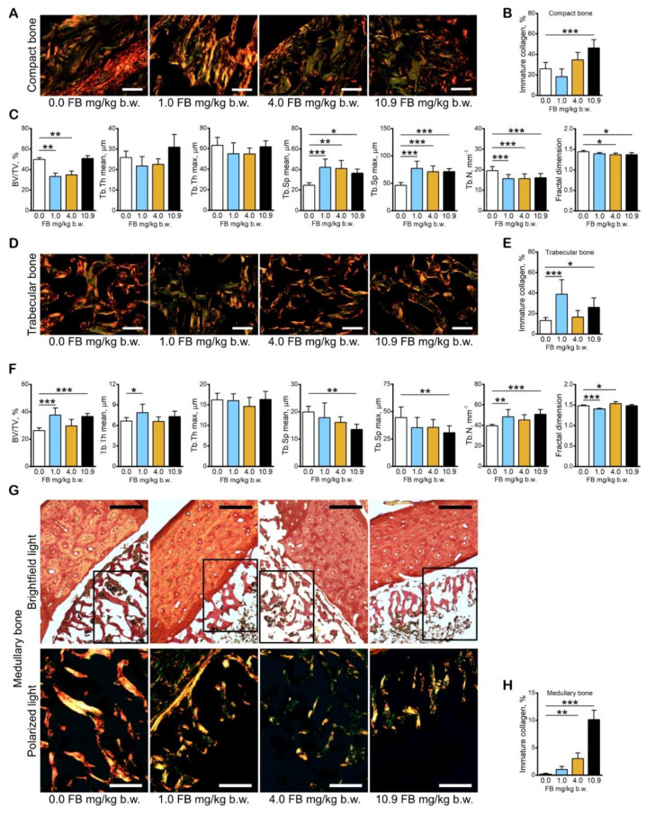
Figure 6.
The effect of fumonisin intoxication on the bone content of immature collagen and bone histomorphometry in pre-laying hens. (A) Representative pictures of the distribution of thin, immature collagen fibers in PRS-stained sections of compact bone observed in polarized light; (B) content of thin, immature collagen fibers in compact bone; (C) histomorphometric analysis of trabecular bone; (D) representative pictures of the distribution of thin, immature collagen fibers in PRS-stained sections of trabecular bone observed in polarized light; (E) content of thin, immature collagen fibers in trabecular bone; (F) histomorphometric analysis of medullary bone; (G) representative pictures of the distribution of thin, immature collagen fibers in PRS-stained sections of medullary bone observed in brightfield light (the marked sections are presented in bottom panels in polarized light); (H) content of thin, immature collagen fibers in medullary bone. Scale bars: A and C: 20 µm; G: 200 µm upper panels and 100 µm bottom panels. B, C, E, F, H: data are expressed as mean ± standard error (n = 8 in each group). Significance was established for fumonisin-intoxicated groups versus the control group (no FB) using a one-way ANOVA followed by a Dunnett’s post-hoc test (normally distributed data) or a Kruskal–Wallis ANOVA with a Dunn’s post-hoc test (for pairwise comparisons with at least one non-normally distributed dataset); * p < 0.05; ** p < 0.01; *** p < 0.001.

